How 3D Printing Helps Scientists Study Plants
Published on
• Last modified on
3D printing isn’t just a novelty process at school science fairs and trade show conferences anymore. It’s everywhere and can make almost everything.
Toys, machine parts, medical implants, footwear, and even a house are being produced with 3D printers.
Now, thanks to researchers at North Carolina State University, plant cells have been 3D bioprinted as well.
“Instead of 3D printing ink or plastic, we use ‘bioink,’ or living plant cells,” said Lisa Van den Broeck, in a release about the study. “The mechanics are the same in both processes with a few notable differences for plant cells: an ultraviolet filter used to keep the environment sterile and multiple print heads-rather than just one-to print different bioinks simultaneously.”
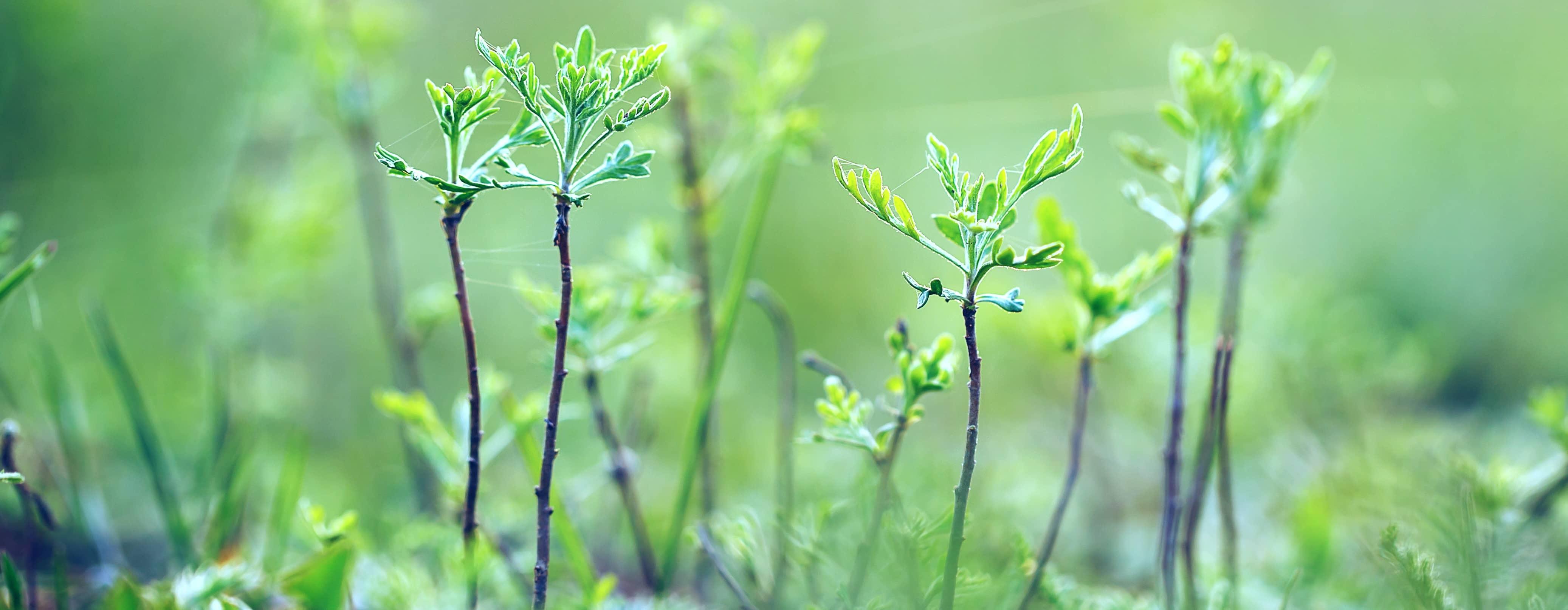
In the study from North Carolina State University, researchers bioprinted cell types from the model plant Arabidopsis Thaliana and from soybeans.
They wanted to see if plant cells would live and if so, how long they would survive after being bioprinted.
They also wanted to demonstrate a reproducible way of studying cellular communication and to examine how plant cells acquire and change their identity and function.
Learning more about how plant cells communicate with each other and with their environment is key to understanding more about plant cell functions. This could ultimately lead to creating better crop varieties and optimal growing environments.
“A plant root has a lot of different cell types with specialized functions,” continued Van den Broeck. “There are also different sets of genes being expressed; some are cell-specific. We wanted to know what happens after you bioprint live cells and place them into an environment that you design. Are they alive and doing what they should be doing?”
Live plant cells without cell walls, or protoplasts, were bioprinted along with nutrients, growth hormones and a thickening agent made from seaweed called agarose. You can think of agarose as the mortar between bricks in the wall of a building. It provides cell strength and scaffolding.
It turns out that while you can 3D print living plant cells, you need to give cells a structure. It’s just like in nature.
“We found that it is critical to use proper scaffolding,” said Ross Sozzani, professor of plant and microbial biology at NC State. “When you print the bioink, you need it to be liquid, but when it comes out, it needs to be solid. Mimicking the natural environment helps keep cellular signals and cues occurring as they would in soil.”
The study found that more than half of the 3D bioprinted cells were viable and divided over time to form small cell colonies.
“We expected good viability on the day the cells were bioprinted, but we had never maintained cells past a few hours after bioprinting, so we had no idea what would happen days later,” added Van den Broeck.
While the Arabidopsis root and shoot cells needed different combinations of nutrients and scaffolding for viability, more than 40% of the individual soybean embryonic cells were viable two weeks after bioprinting. They also divided over time.
“This shows that 3D bioprinting can be useful to study cellular regeneration in crop plants,” said Sozzani. “It also shows the potential of using 3D bioprinting to identify the optimal compounds needed to support plant cell viability in a controlled environment.”